Clinical photos of MCC
Photos of Merkel cell carcinomas
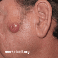
MCC in ear
A Merkel cell carcinoma arising in the ear that was thought to be a squamous cell carcinoma.
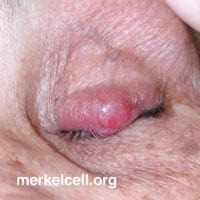
MCC on eye
A Merkel cell carcinoma arising on the eye in an 80 year old woman that was thought to be a stye.
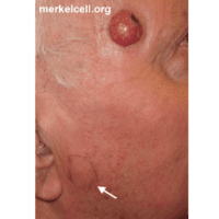
MCC arising on the right temple.
Arrow indicates a firm, enlarged, elevated lymph node (ink shows edges of firmness noted on physical exam), into which MCC tumor cells had spread. Darker area on upper right side of MCC lesion represents site of biopsy.
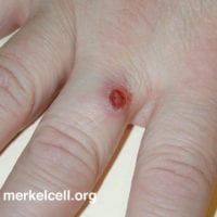
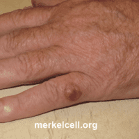
MCC arising on the fifth digit of the left hand.
MCCs are more likely to arise on the left side, likely in part because of greater UV-exposure to the left side when driving.
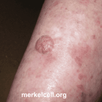
MCC arising on the arm.
The tumor has some features of a wart, but on closer examination the skin surface is quite smooth and blood vessels are enlarged and visible, features that would not be very common for a wart.
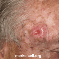
MCC arising on the right temple
The tumor developed in an area of extensive sun damage. This lesion is a Merkel cell carcinoma and squamous carcinoma (SCC) “collision tumor,” meaning the two tumors are directly adjacent to each other. Collision tumors such as this are caused by sunlight and are almost always negative for the Merkel cell polyomavirus.
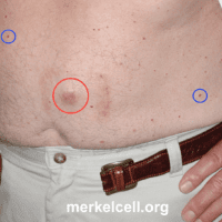
MCC arising on the abdomen (red circle).
The abdomen is a relatively sun protected area but MCC can develop in these areas also. The square-shaped rash around the tumor is a reaction to a bandage. There are cherry angiomas (blue circles), that are 2-3mm red bumps scattered on the abdomen. Cherry angiomas are common benign skin lesions (blood vessel growths) that are unrelated to the MCC.
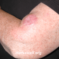
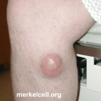
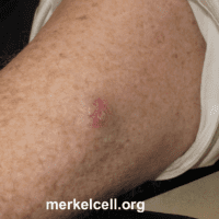
MCC arising on the upper arm.
The lesion was suspected to be a cyst and the MCC diagnosis was a surprise to the patient and physician.

